| Table of Contents |  |
|
Original Article
| ||||||
| A prospective study to evaluate the outcome of operative treatment of patients with intertrochanteric fracture of femur with cephalomedullary nail and dynamic hip screw device | ||||||
| Riyaz N. N.1, Nithin S.2 | ||||||
|
1Associate Professor, Department of Orthopaedics, Academy of Medical Sciences, Pariyaram Medical College Hospital, Kannur-670503, Kerala; India.
2Resident, Department of Orthopaedics, Academy of Medical Sciences, Pariyaram Medical College Hospital, Kannur-670503, Kerala; India. | ||||||
| ||||||
|
[HTML Abstract]
[PDF Full Text]
[Print This Article]
[Similar article in Pumed] [Similar article in Google Scholar] |


| How to cite this article |
| Riyaz NN, Nithin S. A prospective study to evaluate the outcome of operative treatment of patients with intertrochanteric fracture of femur with cephalomedullary nail and dynamic hip screw device. Edorium J Orthop 2015;1:1–7. |
|
Abstract
|
|
Introduction:
It is universally agreed that the treatment of intertrochanteric fractures is stable internal fixation as early as possible. Cephalomedullary nail is gaining popularity than dynamic hip screw over the last few years. The aim of this prospective study was to analyze the patient outcome after dynamic hip screw (DHS) and cephalomedullary nail (CMN) fixation.
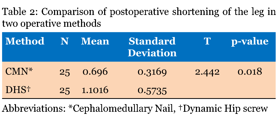
Methods: The study was conducted in Yenepoya Medical College Hospital in Mangalore from 2007 to 2009. Study included 50 patients with intertrochanteric fractures treated with DHS and CMN. Sample size, 25 patients in each group were selected. Parameters compared include, duration of hospital stay, duration of surgery, fracture union, complications and functional outcome. DHS and CMN groups did not differ significantly with respect to age (p = 0.612), complication, pre and postoperative mobility score (p = 0.164), duration of union (p = 0.734) and functional outcome of the stable fractures. Patients with CMN required shorter incision (mean = 5.96 cm) when compared with DHS (mean = 14.04 cm). Surgical time with DHS was more (mean = 98.20 min) when compared with CMN (mean = 53.00 min). Patients treated with CMN experienced less postoperative pain when compared to DHS. Significantly less limb length shortening was seen with CMN (mean = 0.696 cm) as compared to DHS (mean = 1.016 cm). Patients in CMN group with unstable fracture shows better functional outcome when compared to patients with DHS. Conclusion: In stable intertrochanteric fractures, both the CMN and DHS have similar outcomes and for unstable intertrochanteric fractures CMN have shown better functional outcome when compared to DHS. | |
|
Keywords:
Cephalomedullary nail, Dynamic hip screw, Intertrochanteric fractures, Internal fixation, Proximal femoral nail
| |
|
Introduction
| ||||||
|
The special problems of injury and disease in the aged are of growing importance for medical and social reasons. The percentage of old people among our population is increasing and this increase will continue as mortality rate is coming down because of better health care and other reasons. Fractures of trochanteric region of femur are more common in this age group and their management has created considerable interest in this century [1]. For many, this fracture is often a terminal event resulting in death due to cardiac, pulmonary or renal complications. Approximately, 29–38% of patients die within one year of an intertrochanteric fracture [2]. It is thus important to strive to improve treatment and to develop better surgical devices. During recent decades comparison between dynamic hip screw (DHS) and cephalomedullary nail (CMN) had been thoroughly assessed. The DHS has got advantage on implantation where has CMN is technically demanding in implantation. Many studies have shown advantage of CMN over DHS with regards to smaller incision, lesser operating time, and stability. According to Rosenblum et al., intramedullary devices provided three point fixations, a more efficient load transfer due to its medial location with a shorter lever arm and hence, less tensile strain on the implant, reducing the risk of mechanical failure [3]. Baumgaertner et al. concluded that fractures stabilized by an intramedullary hip screw required 10% less operative time and had significantly less blood loss (245 cc v/s 340 cc) than those stabilized with the sliding hip screw [4]. A prospective randomized study of 100 intertrochanteric fractures conducted by Hardy et al. showed that the mean mobility score was significantly greater at one month and three months for the patients who had an intramedullary nail and was had significantly less sliding of the lag screw and subsequent shortening of the limb as compared to those treated with dynamic hip screw device [5]. Few studies have also shown no difference and advantage of DHS over CMN with respect to outcome of the patient. Bridle et al. prospectively compared fixation of 100 intertrochanteric fractures treated randomly by either DHS or intra medullary device like the proximal femoral nail and found no difference in operating time, blood loss, wound complications and final mobility [6]. Kukla et al. recommended the use of intramedullary device only for unstable peritrochanteric fractures after studying 1000 consecutive patients treated with this device between 1992 and 1998 [7]. The main aim of the present study was to evaluate the outcome of operative treatment of intertrochanteric fracture of femur with the CMN and DHS device, with respect to:
| ||||||
|
Materials and Methods
| ||||||
|
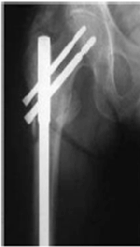
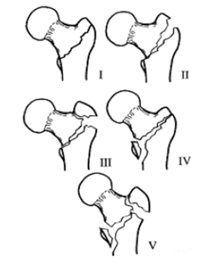
Sixty-four patients suffering from intertrochanteric hip fractures were treated at Yenepoya Medical College Hospital, Derlakatte, Mangalore from July 2007 to August 2009. The intertrochanteric fracture taken for study were fresh fractures which were studied prospectively after taking due consent. The prospective study compared the outcome of operative treatment of intertrochanteric fracture of femur with CMN (Figure 1) and DHS (Figure 2) device. Patients were put into two groups based on the method of surgery. The outcome of the patient in two groups was decided based on the duration of hospital stay, duration of surgery, fracture union, functional outcome and complications. Fractures in both the groups were classified into stable and unstable based on Jensen and Michaelsen's modification of Evans Classification (Figure 3) and was compared based on the functional outcome of the patient. All the patients were initially evaluated as to their general condition, hydration and corrective measures were undertaken. Antero-posterior radiograph of the affected hips were taken. The patients were then put on skeletal traction or skin traction over a Bohler-Braun frame. No open fractures were encountered in this series. Patients were taken up for surgery as soon as their general condition permitted. Adequate blood transfusion, thromboprophylaxis and other supportive measures were given depending on the preoperative condition of the patient and also post surgery based on the blood loss during surgery. Surgery (DHS or CMN) was decided, depending on the morphology of fracture, affordability of the patients, facility and experience of the operating surgeons. Of the 64 patients in the study, 35 were treated with DHS and 29 with CMN. Fourteen patients lost for the follow- up. Two patients associated with ipsilateral proximal Humerus fractures were encountered in the study. Out of 64 fractures, 55 fractures reduced by closed method and 9 fractures reduced by open method. The length of the incision and duration of surgery was recorded intraoperatively. There was no defined postoperative patient protocol, but all patients were given postoperative injectable antibiotics for 3–5 days followed by oral antibiotics for next 1 week and deep venous thrombosis prophylaxis (squeezing calf muscles, in bed mobilization, static quadriceps strengthening exercises). Patients were allowed to sit up in bed on the second postoperative day. Static quadriceps exercises where started on the second and third postoperative day. Drain was removed after 2nd postoperative day. Sutures were removed after 10–14 days. Patients were mobilized non-weight bearing as soon as the pain or general condition permitted. Weight bearing was commenced depending upon the stability of the fracture and adequacy of fixation, delaying it for six weeks, for patients with unstable or inadequate fixation. All the patients were followed-up for a period of minimum six months and maximum of one year.Functional assessment was based on Jensen and Michaelsen's modification of Evans Classification [8] [9] (Figure 3). The outcome was assessed based on the postoperative pain, walking ability, hip joint range of motion, and limb length shortening as follows: This study involves patient's manipulation and is the method of choice advocated by authors in standard books and international journals. So ethical clearance was obtained prior to the study from the ethical committee of the institution and prior consent was obtained from each of the participants. The collective data was analyzed by Student t-test and chi-square test using SPSS software to evaluate the results. | ||||||
| ||||||
| ||||||
|
| ||||||
| ||||||
|
Results | ||||||
|
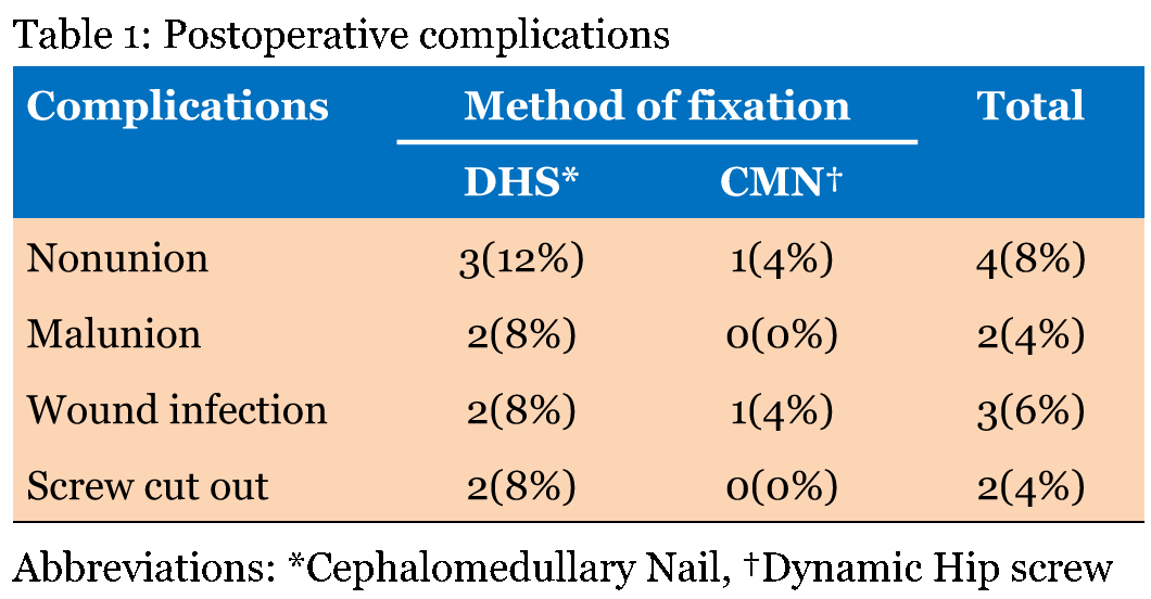
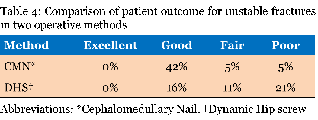
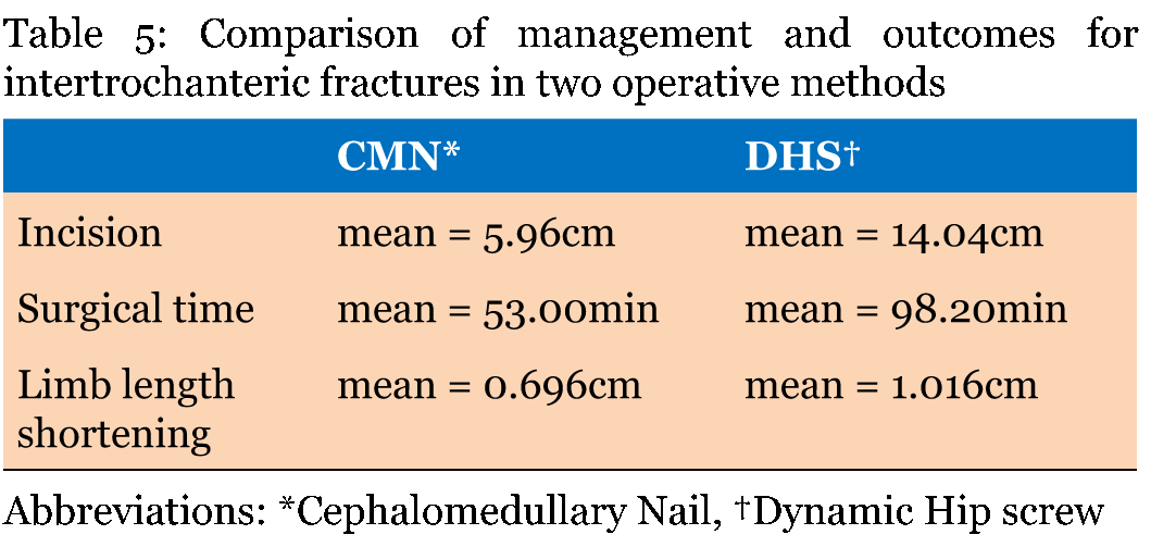
Of the 64 patients in the study, 35 were treated with DHS and 29 with CMN. Fourteen patients lost for the follow up. Two patients with associated ipsilateral proximal Humerus fractures were encountered in the study. Out of 64 fractures, 55 fractures reduced by closed method and 9 fractures reduced by open method. Variables considered for the study were age, length of incision, duration of surgery, postoperative pain, preoperative and postoperative mobility score, postoperative shortening, postoperative range of motion, duration of union and type of fracture. Present study assessed the outcome of the patients who were treated with DHS and CMN with respect to the above mentioned variables. There was no significant association (p = 0.612) between the age and functional outcome of the patients in two groups. With regards to the length of incision there was significant difference (p = 0.0005) between the two groups. Patients treated with CMN require short incision (mean = 5.96 cm) when compared to patients treated with DHS (mean = 14.04 cm). Duration of surgery was statistically significant (p = <0.0005) in two groups. Surgical time for patients with DHS was more (mean = 98.20 min) when compared to patients with CMN (mean = 53.00 min). Presence of pain during mobility is direct indicator of the functional outcome of the patient. Patients were revived monthly and pain score during mobility was obtained during each visit. Figure 4 shows the presence of pain during mobility had significant association (χ2 = 17.943) between the two groups at sixth month of follow-up. Patients treated with CMN experienced less postoperative pain when compared to patients treated with DHS. Of 50 patients 11 patients encountered postoperative complications (Table 1) which include nonunion, malunion, wound infection and screw cutout. There was no significant difference between the two groups with regards to post-operative complications. Preoperative and postoperative mobility score between two groups were compared at fourth month of follow-up. Analysis showed no significant difference (p = 0.164) between the two groups. Postoperative shortening of the operated limb is an indicator of implant failure or success. Significantly (p = 0.018) less limb length shortening was seen in CMN group (mean = 0.696 cm) as compared to the DHS group (mean = 1.016 cm) (Table 2). With regards to postoperative range of motion there was no significant (p = 0.147) difference in two groups. Results showed no significant difference (p = 0.734) between the duration of union of fractured bones between two groups at one year of follow-up. Fractures in both groups were classified into stable and unstable based on Jensen and Michaelsen's modification of Evans classification. Functional outcome of the patients were established in both groups based on the fracture classification (Table 3) and (Table 4) . The results showed no significant difference in both groups for stable fracture, but for unstable fracture the results were highly significant (p < 0.0005) for both groups. This indicates that the patients in CMN group with unstable fracture shows better functional outcome when compared to patients in DHS group (Table 5) . In CMN group, among 15 stable fractures 80% had good to excellent results and in DHS group among 16 stable fractures 87.5% had good to excellent results. In CMN group, among 10 unstable fractures 80% had good to excellent results and in DHS group among 9 unstable fractures 33% had good to excellent results. This indicates that the patients in CMN group with unstable fracture shows better functional outcome when compared to patients in DHS group. | ||||||
| ||||||
| ||||||
| ||||||
| ||||||
| ||||||
|
| ||||||
| ||||||
|
Discussion
| ||||||
|
Intertrochanteric fracture is one of the most common fractures of the hip especially in the elderly with porotic bones, usually due to low-energy trauma like simple falls. The incidence of intertrochanteric fracture is rising because of increasing number of senior citizens with osteoporosis. By 2040, the incidence is estimated to be doubled. In India, the figures may be much more. Problems of these fractures are [1] associated with substantial morbidity and mortality [2] malunion [3] implant failure, cutout of head, and penetration into hip [4] great financial burden to the family [5] associated medical problem like diabetes, hypertension [10]. It is thus important to strive to improve treatment and to develop better surgical devices. The new treatment modalities must prove their usefulness and superiority over the old methods, even in terms of functional outcome, which is an aspect lacking from most of the earlier comparisons between CMN and DHS. Present study was a prospective study comparing CMN and DHS by focusing on duration of hospital stay, duration of surgery, fracture union, complications and functional outcome. In the present study, there was no significant difference between the age and the type of fixation. Patient treated with CMN require short incision when compared to patients treated with DHS. This finding was supported by the study conducted by Leung et al. [11] in which the average length of incision was 15.7 cm in DHS group and 8.9 cm in CMN group. Surgical time for patients with DHS was more (mean = 98.20 min) when compared to patients with CMN (mean = 53.00 min). According to Adams et al. [12] average duration of surgery was 61.3 minutes for DHS and 55.4 minutes for CMN group. This difference was mainly because CMN requires short incision when compared to DHS. Patients treated with CMN experienced less post-operative pain when compared to patients treated with DHS. Saudan et al. [13] found that the amount of persistent pain was similar in both CMN and DHS group. Patient treated with CMN experienced less postoperative pain in the present study was due to smaller incision length. There will be less blood loss and minimal soft tissue injury which will decrease the pain during mobility postoperatively in short incisions. Of 50 patients 11 patient encountered postoperative complications which include nonunion, malunion, wound infection and screw cutout. There was no significant difference between the two groups with regards to postoperative complications. The finding was supported by Menezes et al. [14] and Saudan et al. [15] in which there was no significance between the type of fixation and the occurrence of postoperative complications. Lower complication rate was evident because of the strict aseptic techniques, proper preoperative planning, effective rehabilitation process and availability of expertise surgeons. Patients in our study treated with CMN regained their pre-injury walking ability at fourth month significantly more often than those treated with DHS. In our series, only 6 of the 25 patients (24%) in the DHS group regained their pre-injury mobility level as compared to 13 of the 25 patients (52%) in the CMN group at the fourth month of follow-up. Similar findings were also seen in a series by Pajarinen et al. [15] . Reason for this difference might be the significantly greater impaction of the fracture in the DHS group with shortening of the proximal femur, thus altering the biomechanics of the hip and preventing restoration of the ability to walk. Moreover, the lack of compression in the CMN group did not seem to interfere with the healing of the fracture. Significantly (p = 0.018) less limb length shortening was seen in CMN group (mean = 0.696 cm) as compared to the DHS group (mean = 1.016 cm). According to Leung et al. [11] less limb length shortening was seen in the DHS group as compared to the CMN group with a mean of 2 cm and 3 cm, respectively. This could be due to the increased sliding of the lag screw in the DHS group, allowing greater fracture impact ion, as compared to the CMN. Patients treated with CMN recovered 73% of their hip range of movement as compared to those treated with DHS who recovered only 68.4% of their hip range of movement. Postoperative range of motion was similar in both groups. According to Leung et al. [11] patients treated with CMN recovered 46.9% of their hip range of movement as compared to those treated with DHS who recovered only 18.09% of their hip range of movement. In our series, all the fractures united at third month time except 3 in DHS and 1 in CMN group which went for nonunion. According to Cleveland et al. [16] all fractures united at an average time of 4.5 months. There is no significant difference in the fracture union in both the groups. A study conducted by Madsen et al. [17] indicates better patient functional outcome for unstable fracture with the use of trochanteric stabilizing plate when compared to DHS. This significant difference in unstable fracture is mainly because the DHS method is not advisable for unstable comminuted fracture and in cases where lateral cortex is not intact. Present study is only comparing the difference between DHS and CMN with respect to the outcome of the patient. It is not considering the various sub types of cephalomedullary nail including Gamma nail, Proximal Femoral nail, Trochanteric Femoral nail and Recon nails. Further research can be conducted on comparison of different types of cephalomedullary nail with respect to the patient outcome. Researcher has only taken fresh cases in the present study, so further study can be conducted comparing the CMN and DHS on non union intertrochanteric fractures with regards to patient outcome. It is concluded that both methods are useful in the treatment of intertrochanteric femoral fracture, although the results were in favor of cephalomedullary nail with respect to the length of incision, duration of surgery, presence of postoperative pain, postoperative limb shortening and functional outcome of unstable fractures. | ||||||
|
Conclusion
| ||||||
|
Proximal femoral fractures are usually treated surgically. In the last decade, extramedullary methods of fixation with various angular plates or with a compression hip screw with a plate are more and more replaced by newer intramedullary techniques because of their advantages: the surgical procedure is faster, the blood loss is less, the bone healing mainly remains in the reduced position with a biomechanically strong fixation, that allows earlier weight bearing on the bone with less local and systemic complications. Osteosynthesis with the cephalomedullary nail offers the advantages of high rotational stability of the head-neck fragment. Cephalomedullary nail has the advantage of collapse at fracture site and is biomechanically sound as it is an intramedullary device. Postoperatively, early mobilization can be begun as the fixation is rigid. Cephalomedullary nail being an intramedullary load sharing device offers better biomechanical stability. In stable intertrochanteric fractures, both the dynamic hip screw (DHS) and cephalomedullary nail (CMN) have similar outcomes and for unstable intertrochanteric fractures CMN have shown better functional outcome when compared to DHS. | ||||||
|
Acknowledgements
| ||||||
|
We are thankful to Late Prof.M. Sudhakar Shetty for his valuable suggestions and timely advices. | ||||||
|
References
| ||||||
| ||||||


|
[HTML Abstract]
[PDF Full Text]
|
|
Author Contributions:
Riyaz N. N. – Substantial contributions to conception and design, Acquisition of data, Analysis and interpretation of data, Drafting the article, Revising it critically for important intellectual content, Final approval of the version to be published Nithin S. – Substantial contributions to conception and design, Acquisition of data, Analysis and interpretation of data, Drafting the article, Revising it critically for important intellectual content, Final approval of the version to be published |
|
Guarantor of submission
The corresponding author is the guarantor of submission. |
|
Source of support
None |
|
Conflict of interest
Authors declare no conflict of interest. |
|
Copyright
© 2015 Riyaz N. N. et al. This article is distributed under the terms of Creative Commons Attribution License which permits unrestricted use, distribution and reproduction in any medium provided the original author(s) and original publisher are properly credited. Please see the copyright policy on the journal website for more information. |
|
|