 |
|
Case Report
| ||||||
| A novel technique for treatment of a pathological fracture in a distal humerus using combined interlocking custom shoulder and elbow arthroplasty | ||||||
| Mohamed Hafez1, Ian A Trail2 | ||||||
|
1MSc, MRCS, FRCS Upper limb fellow, upper limb unit, Wrightington, Wigan, UK 2MD FRCS, Consultant upper limb upper limb unit, Wrightington, Wigan, UK | ||||||
| ||||||
|
[HTML Abstract]
[PDF Full Text]
[Print This Article]
[Similar article in Pumed] [Similar article in Google Scholar] |


| How to cite this article |
| Hafez M, Trail IA. A novel technique for treatment of a pathological fracture in a distal humerus using combined interlocking custom shoulder and elbow arthroplasty. Edorium J Orthop 2017;3:13–16. |
|
ABSTRACT
|
|
There is no uniform plan for the management of patients with metastatic lesions within the humerus. Any plan has to be tailored to each individual patient due to a variety of factors, including the clinical presentation, stage of disease, general condition, together with the skills and resources of the clinical team. Satisfactory management requires a multidisciplinary team approach, including an orthopedic surgeon, medical oncologist, radiation oncologist, pathologist and other support services. We report a case of a multiple tumor metastasis within the humerus, complicated by a pathological fracture of the distal humerus, treated with a combined ipsilateral shoulder and elbow arthroplasty using a custom interlocking prosthesis. The postoperative course was uneventful and the patient showed excellent functional and clinical improvement. | |
|
Keywords:
Combined shoulder elbow replacement, Humerus tumors, Pathological fracture
| |
|
INTRODUCTION
| ||||||
|
Metastatic lesions are the most common malignant neoplasm in bone [1]. According to Clain proximal humerus comes only after femur in the frequency of secondary tumor spread [2]. Breast is the most common primary focus to cause pathological fracture in humerus (35–41%) [3]. It is well documented that the improvement of medical management of primary tumors has resulted in a significant increase in life expectancy. Patients with metastatic breast cancers have a mean survival of 28 months [1]. The decision whether to proceed with surgery or not is usually made following consultation between the patient, oncologist and surgeon. Sources were retrieved via a PubMed search. Search terms such as plica syndrome, synovial plicae, and synovial joint morphogenesis were utilized to identify relevant sources. These searches yielded 142, 112, and 446 results, respectively. Sources were included if they were considered relevant to the current review article, which was determined by analyzing the title or abstract of the source. Additionally, sources that were cited in other review articles were retrieved if they were relevant to the current review article and they were not retrieved in the original PubMed search. The surgical options for treatment of pathological fractures within the distal humerus include open reduction and internal fixation, using compression plate with or without poly methylmethacylate cement (PMMA) or endoprosthetic replacement of the humerus. For proximal humerus metastasis, surgical management includes humeral replacement or forequarter amputation. Humeral replacement has always been criticized as being no more than a spacer than a true articulating construct [1]. There is no doubt, however, that salvage surgeries are more acceptable to patients and the function is markedly superior to amputation. | ||||||
|
CASE REPORT
| ||||||
|
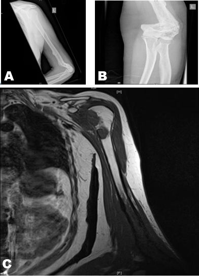
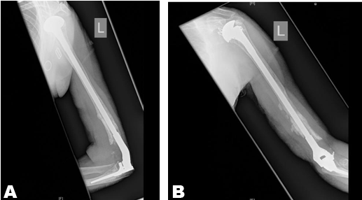
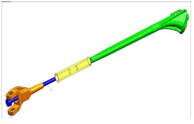
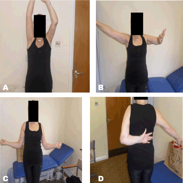
A 55-year-old Jehovah’s witnesses lady was referred to the senior author (IAT) with a pathological fracture at distal humerus and multiple metastatic lesions of proximal humerus secondary to breast cancer. Magnetic resonance imaging scan showed a non-union of a pathological fracture with extensive metastatic involvement of the distal humerus as well as multiple large metastases in the proximal humerus. Following consideration of the history, physical examination, radiographs and discussion with the medical oncologist, the senior author concluded that a combined linked shoulder and elbow arthroplasty was the appropriate treatment option. At the time of surgery, after appropriate preoperative assessment and planning, the patient was anesthetized in a supine position. At the shoulder, a deltopectoral approach was used to insert a hemiarthroplasty. The latter was cemented in situ and the wound closed in layers. The patient was then repositioned in a lateral decubitus and a posterior approach to the elbow and distal humerus undertaken. Following resection of the radial head, the ulna was prepared. After the humeral component was inserted through the distal fragment, this was then secured to the tip of the proximal stem, using a locking mechanism. At that time great care was taken to make sure that the alignment was appropriate. The ulnar component was then cemented in place and the two components of elbow replacement connected. Thereafter, the wound was closed in layers. The implants used were a Depuy custom modified global humeral component, using a shoulder eccentric head and an Acclaim elbow used in a format. Operation time was 2 hours and 5 minutes. No blood transfusion was necessary during or postoperatively. The patient maintaining a hemoglobin level above 8.8 g/dL during her recovery. Postoperatively, the arm was immobilized for three weeks in a splint. Thereafter, active mobilization of the shoulder and elbow was instigated and the patient made an uneventful recovery. During the postoperative phase, she received a course of irradiation to the arm. At the time of review, one year after surgery, she was asymptomatic and her limb maintained an adequate range of motion and function. She had a Constant Score of 76, quick DASH of 18.2, ASES score of 90 and MEPS 80. Radiographs taken showed good position of the prosthesis (Figure 1) (Figure 2) (Figure 3) (Figure 4). | ||||||
|
| ||||||
| ||||||
| ||||||
| ||||||
|
DISCUSSION
| ||||||
|
Metastatic tumors around the elbow are rare [4]. Usually, patients present with either pain or a pathological fracture. The surgical management of pathological fractures around the elbow is challenging, due to the uncommon nature of the condition and the lack of guidance in literature. The aim of treatment is to achieve pain relief and to restore maximum motion and stability, thus providing a functional upper limb. With advances in imaging, staging and perioperative oncological management, life expectancy has increased and the debate as to whether salvage or amputate the limb has shifted to the former. Nowadays, reconstruction of the upper limb has become the standard treatment for patients with bone tumors. Compared to the other options of elbow reconstruction after bone resection, prosthetic replacement is the option of choice, in that arthrodesis is poorly tolerated and technically demanding, especially after tumor resection, osteoarticular allografts have a high complication rate and excision arthroplasty is rarely an option, due to the large bone gap [5]. For pathological fractures, open reduction and internal fixation is always a valid option, but it has a high nonunion rate [6]. Athwal et al., reported 20 patients after a Coonard Morey elbow replacement, undertaken for tumors of the distal ulnar. Although 18 patients also had perioperative radiotherapy/chemotherapy, the infection rate was 0%, 45% of patients experienced ulnar or radial nerve complication after surgery and local recurrence was 25%. These results demonstrated that a total elbow replacement gives good pain relief, preserves function and the oncological and non-oncological complications are comparable to other reconstruction techniques. These results demonstrated that within this clinical scenario, total elbow arthroplasty gives good pain relief. The linked elbow implant used in this study can compensate for variable degrees of bone loss, although the survival of the implant depends on many factors. According to Shah et al, these would include the overall alignment of the implant, surgical skills, as well as the quality of the patients’ bone [7][8]. Another major factor is the humeral stem length, an increase from 3.5–7 cm has increased the survival by decreasing humeral loosening Trail el al. [8][9]. The standard method of treatment for the proximal humerus is again to salvage the limb, in that reconstruction following excision of tumors of the proximal humerus gives better function and is more cost effective when compared to amputation [3][10] Allograft, arthrodesis and endoprosthetic replacement of proximal humerus (EPRPH) have all been used [10]. When comparing the shoulder hemiarthroplasty carried out in our patient with the formal EPRPH, it is of note that the level of bone resection was intra-articular and, as such, neither the rotator cuff nor axillary nerve was compromised. This undoubtedly resulted in improved movement, particularly abduction. Periprosthetic fracture is the major implant related complication with ipsilateral shoulder and elbow replacements [4][11]. Many studies have tried to find a solution for this problem. One approach suggested leaving a gap of more than 6 cm between the tips of the prosthesis, [4] another one suggested minimizing this gap [11]. One more study suggested filling the space between the implants with cement [4]. Plausinis et al. studied the mechanics behind these approaches and showed that the gap between the tips of implants whether it was 5-30-60 mm did not affect the stress concentration at the tips of implants. In addition, they also showed that filling the space between the implants with cement did not significantly affect fracture rate, as only 3% of the bending forces were transmitted across the cement [12]. Stress shielding is another complication of arthroplasty that can lead to periprosthetic fractures[11][13]. The incidence of stress shielding increase with increasing the stiffness of the implant. In our case, although there is an increased risk of developing stress shielding, current radiological evaluation does not show any evidence of progressive cortical thinning or impending fractures. | ||||||
|
CONCLUSION
| ||||||
|
In conclusion, because of the reported high risk of periprosthetic fractures after ipsilateral shoulder and elbow replacement, we used a linked prosthesis, spanning the humerus with an internal strut, allowing function to be maintained. | ||||||
|
REFERENCES
| ||||||
| ||||||

|
[HTML Abstract]
[PDF Full Text]
|
|
Author Contributions
Mohamed Hafez – Substantial contributions to conception and design, Acquisition of data, Analysis and interpretation of data, Drafting the article, Revising it critically for important intellectual content, Final approval of the version to be published Ian A. Trail – Analysis and interpretation of data, Revising it critically for important intellectual content, Final approval of the version to be published |
|
Guarantor of submission
The corresponding author is the guarantor of submission. |
|
Source of support
None |
|
Conflict of interest
Authors declare no conflict of interest. |
|
Copyright
© 2017 Mohamed Hafez et al. This article is distributed under the terms of Creative Commons Attribution License which permits unrestricted use, distribution and reproduction in any medium provided the original author(s) and original publisher are properly credited. Please see the copyright policy on the journal website for more information. |
|
|