 |
Editorial
Morel Lavallee lesion with pelvic and acetabular fractures; Operate or wait?
1 Orthopedic and Traumatology Department, Sohag Faculty of Medicine, Sohag University, Sohag, Egypt
Address correspondence to:
Hossam Hosny
Sohag University, Sohag 82648,
Egypt
Message to Corresponding Author
Article ID: 100013O03HH2019
Access full text article on other devices

Access PDF of article on other devices


How to cite this article
Hosny H. Morel Lavallee lesion with pelvic and acetabular fractures; Operate or wait? Edorium J Orthop 2019;5:100013O03HH2019.ABSTRACT
No Abstract
Keywords: Acetabular fractures, Morel lavallee lesion, Pelvic fractures, Soft tissue injury, Pelvic and acetabular fracture, Wound healing
Editorial
Morel-Lavallee lesion was described by French physician Maurice Morel-Lavallee in the year 1853 [1]. It is a clinical sign that occurs after a major trauma resulting in abrupt separation of the skin and subcutaneous tissue from the underlying fascia. Thus, a space will be created between the fascia overlying the muscle layer and layer of the skin and subcutaneous tissue. The space will be filled by a serosanguinous fluid, blood and necrotic fatty tissue. It is a closed degloving soft tissue injury. The lesion is commonly associated with pelvic and acetabular fractures. Letournel and Judet were the first to use the term "Morel-Lavallee lesion" in the description of such lesion over an acetabular fracture. The trochanteric region and proximal thigh is the most frequent sites for Morel Lavallee lesion [2]. It may occur else in the trunk, proximal humerus, prepatellar and scapular region. The lesion may not be associated with a fracture and occurs after trauma to the affected site. Morel Lavallee lesion usually occurs within hours or it may be delayed up to several months after trauma insult. Obese individuals are considered as a high risk for Morel Lavallee lesion and diagnosis of the lesion in these patients may be delayed up to many days.
DIAGNOSIS OF MOREL LAVALLEE LESION
Clinical examination
The patient had a history of trauma and severe pain at the affected region. On inspection, there is a contusion marks over the affected region and discoloration of the skin. On palpation, a painful fluctuant swelling might be palpated with severe tenderness at the affected region.
Ultrasonographical evaluation
Ultrasound will show massive fluid collection in the subcutaneous region.
Computed Tomography (CT)
Computed Tomography (CT) scan will show a cavity formation with hypodense collection within the subcutaneous region associated with fatty density within the lesion.
Magnetic Resonance Imaging (MRI)
MRI is the most accurate method to diagnose Morel Lavallee lesion. It will show fluid collection beneath the skin and subcutaneous tissue and overlying the muscle layer. The collection appears hypo-intense with hyperintense (fat) strands on T1-weighted (T1W) images and hyperintense on T2-weighted (T2W) images.
According to MRI findings, Mellado and Bencardino classified Morel Lavelle lesions into six types [3]:
Type I: Laminar shaped and seroma-like with increased T2 signal.
Type II: Oval-shape that resembling a subacute hematoma with increased T1 and T2 signal; thick capsule and variable enhancement.
Type III: Oval shaped just like a chronic organizing hematoma; thick capsule and internal/peripheral enhancement.
Type IV: Linear; resembles a closed laceration with hypointense T1 signal and hyperintense T2 signal; no capsule and variable enhancement.
Type V: Pseudonodular with a round shape, variable T1 and T2 signal, a thin or thick capsule, internal/ peripheral enhancement.
Type VI: Infected with variable T1 and T2 signal; variable sinus tract formation, a thick capsule, and internal/peripheral enhancement.
Differential Diagnosis of Morel Lavallee Lesion
The lesion mimics hemangioma, subcutaneous hematoma, soft tissue sarcoma and fat necrosis.However, the location, history of trauma, clinical examination and radiographic evaluation (Ultrasonography, CT Scan and MRI) can distinguish Morel Lavallee lesion from other lesions.
TREATMENT
Treatment of Morel Lavallee lesion depends on many factors as size, site and its relation to the surgical approach. Small lesions can be treated conservatively by applying a compression bandage. If these lesions do not resolve spontaneously, it will need aggressive treatment by surgical debridement. Large lesions can be treated with early percutaneous drainage, debridement, irrigation, injection of sclerosing agents such as tetracycline and suction drainage.
Management of acetabular and pelvic fractures associated with Morel Lavallee Lesion
As Morel Lavallee lesion is more common with pelvic and acetabular fractures. The decision to go for surgical treatment of these fractures is still controversy for all orthopedic surgeons.
Steiner et al in their study of twenty patients with Morel Lavallee lesion and associated acetabular and pelvic fractures, advised open debridement of the lesion during or before osteosynthesis if the lesion is lying at the site of surgical approach. If the lesion is away, they used an incision to debride the lesion and another incision to fix the fracture [4].
Tseng et al in their study of nineteen patients with Morel Lavallee lesion, fifteen patients had associated pelvic and acetabular fractures. Nine patients had a pelvic fracture while six patients had acetabular fractures. Among the nine patients, seven patients in whom a pelvic fracture was treated surgically had percutaneous fixation of the posterior part of the pelvic ring. The treatment of the Morel Lavallee lesion was also done during the same operative setting and none of them had infectious complications or sustained skin loss. Fixation of the remaining two pelvic fractures and the six acetabular fractures was delayed until at least twenty-four hours after the drain was removed after five to fifteen days after debridement.
Three patients who had acetabular fractures had a surgical incision directly through the lesion (lateral approach to the hip and Kocher-Langenbeck approach). The other three patients with acetabular fractures had a surgical incision away from the lesion (ilioinguinal approach and anterior approach to the pelvis) [5].
The decision for surgical intervention in cases with acetabular and pelvic fractures associated with Morel Lavallee lesion should depend on many factors. Most of cases that had Morel Lavallee lesion must have a special care as these cases associated with high morbidity and mortality rates.
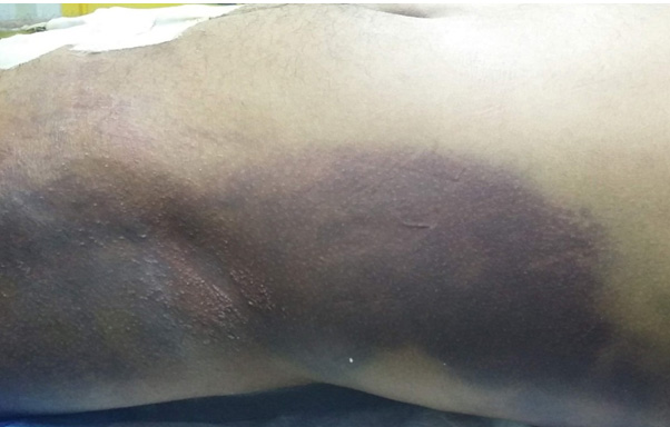
In cases with small lesions that associated with discoloration and contusion marks over the skin without fluctuant sensation on palpation, surgery is considered safe even if the lesion is overlying the region of surgical approach with caution to decrease the operative time and preferring the percutaneous technique and this must be followed by strong broad-spectrum antibiotics and daily close follow up and dressing of the wound. Evacuation of the hematoma if present through the percutaneous technique is essential and beneficial to guard against bacterial colonization (Figure 1).
In cases with moderate sized lesions, surgical intervention to fix the underlying fracture depends on the proximity of the lesion to the region of surgical approach. If the lesion is away, open debridement of the lesion with an incision a long its whole length and another incision for surgical fixation of the fracture. Repeated debridement is essential to keep the wound clean and prevent bacterial colonization. Percutaneous technique and short operative time are preferred to limit the incidence of complications.
If the lesion is overlying the region of surgical approach, controversy exists. Some surgeons advised one stage open debridement and internal fixation of the fracture from the same incision with applying of vacuum sealing drainage [6]. While others prefer repeated open debridement of lesion and fixation of the fracture later after stabilization of the patient.
In fact, the first choice is preferred in cases with fractures needing urgent surgical fixation as unstable pelvic fractures while the other choice should be the standard to fix pelvic and acetabular fractures in cases with moderate sized lesion for many reasons as the metal itself will increase the incidence of infection even if you had a good debridement of the wound moreover the Morel Lavallee lesion has a progressive and aggressive course that cannot be predicted.
In cases with massive Morel Lavallee lesions, open debridement is the best choice to deal with it. Surgical fixation of the fractures in these patients is not allowed and carries hazardous effects and will increase the morbidity and mortality rates. We should know that Morel Lavallee lesion has unpredictable course with high morbidity and mortality rates.
REFERENCES
1.
Morel-Lavallee VAF. Decollements traumatiques de la peau et des couches sous-jacentes. [Article in French]. Arch Gen Med 1863;1:20–38. 172–200, 300–32.

2.
Letournel E, Judet R. Fractures of the Acetabulum. Berlin, Heidelberg: Springer-Verlag; 2012.
3.
Mellado JM, Bencardino JT. Morel-Lavallée lesion: Review with emphasis on MR imaging. Magn Reson Imaging Clin N Am 2005;13(4):775–82. [CrossRef]
[Pubmed]
4.
Steiner CL, Trentz O, Labler L. Management of Morel-Lavallee lesion associated with pelvic and/or acetabular fractures. Eur J Trauma Emerg Surg 2008;34(6):554–60. [CrossRef]
[Pubmed]
5.
Tseng S, Tornetta P 3rd. Percutaneous management of Morel-Lavallee lesions. J Bone Joint Surg Am 2006;88(1):92–6.
[Pubmed]
6.
Wei D, Wang Y, Yuan J, et at. One-stage operation for pelvis and acetabular fractures combined with Morel-Lavallée injury by internal fixation associated with vacuum sealing drainage. [Article in Chinese]. Zhongguo Xiu Fu Chong Jian Wai Ke Za Zhi 2014;28(1):38–42.
[Pubmed]
SUPPORTING INFORMATION
Author Contributions
Hossam Hosny - Conception of the work, Design of the work, Acquisition of data, Analysis of data, Drafting the work, Revising the work critically for important intellectual content, Final approval of the version to be published, Agree to be accountable for all aspects of the work in ensuring that questions related to the accuracy or integrity of any part of the work are appropriately investigated and resolved.
Guaranter of SubmissionThe corresponding author is the guarantor of submission.
Source of SupportNone
Data AvailabilityAll relevant data are within the paper and its Supporting Information files.
Conflict of InterestAuthors declare no conflict of interest.
Copyright© 2019 Hossam Hosny. This article is distributed under the terms of Creative Commons Attribution License which permits unrestricted use, distribution and reproduction in any medium provided the original author(s) and original publisher are properly credited. Please see the copyright policy on the journal website for more information.


